Shoulder
Radiographic Anatomy of the Shoulder
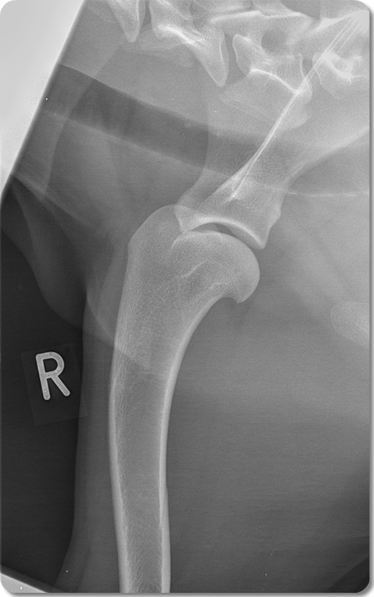
Medio-lateral Projection
Click on the circles to see the answer
Glenoid Cavity of Scapula
Spine of Scapula
Greater Tubercle of Humerus
Insertion of the m. supraspinatus and the partial insertion of the m. pectorals profundus
Clinical relevance:
Tendinopathy of the supraspinatus tendon
Mainly medium to large, middle-aged dogs
Acromion of scapula
Easily palpated in the living animal
Supraglenoid Tubercle of Scapula
Origin of the tendon of the m. biceps brachii.
1st Sternebra (Manubrium)
5th Cervical Vertebra
Head of Humerus
Clinical relevance: Osteochondrosis of the humeral head
Affect young dogs large breed dogs

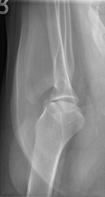
Spine of scapula
Greater tubercle of humerus
Acromion of scapula
Lesser tubercle of humerus
Radiographic Anatomy of the Shoulder
Ventro-dorsal Projection
Click on the circles to see the answer

Computed Tomography Anatomy of the Shoulder
Sagittal plane
Click and drag to go through the study. Position your cursor over the structures to see anatomical labels
- Humerus
- Scapula
- Acromion
- Glenoid Cavity of Scapula
- Supraglenoid Tubercle
- Coracoid Process
- Greater Tubercle of Humerus
- Lesser Tubercle of Humerus
- Head of Humerus
- Spine of Scapula
Computed Tomography Anatomy of the Shoulder
Dorsal Plane
Click and drag to go through the study. Position your cursor over the structures to see anatomical labels
- Scapula
- Humerus
- Acromion
- Glenoid Cavity
- Supraglenoid tubercle
- Coracoid Process
- Greater Tubercle of Humerus
- Lesser Tubercle of Humerus
- Head of Humerus
- Spine of Scapula
Magnetic resonance imaging of the shoulder
3D Reconstruction of the Shoulder
Click and drag to rotate the 3D object
Click on labels to see annotations
Comparative - Equine shoulder
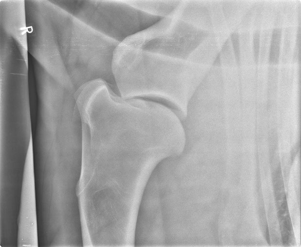
Radiographic Anatomy of the Shoulder
Medio-lateral Projection
Click on the circles to see the answer
Glenoid Cavity of Scapula
Spine of Scapula
Distal end of intermeditate groove
Deltoid tuberosity
Cranial part of the lesser tubercle
5th Cervical Vertebra
Head of Humerus
Floor of the intertubercular groove

Dr Mariano Makara
Dip. ECVDI