Hips
Radiographic Anatomy of the Hips
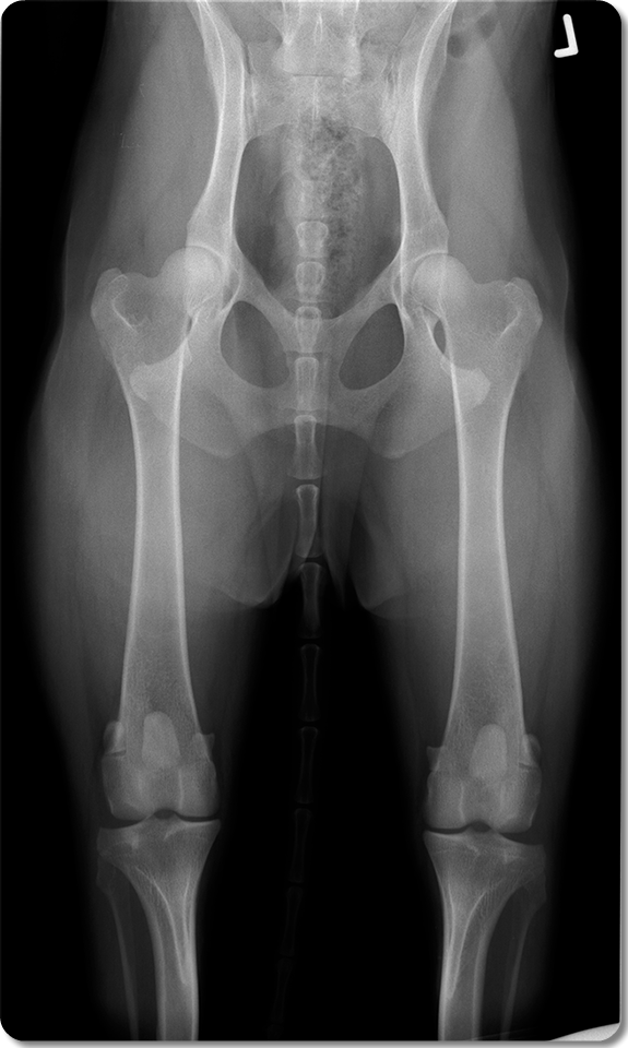
Ventro-dorsal projection
Click on the circles to see the answer
Body of Left Ilium
The ilium is the largest and most cranial of the bones that compose the os coxae. It is divided into the wing and the body
The body is the narrow, more irregular caudal part of the bone. In its caudal end forms the cranial two fifths of the acetabulum. In this cavity it fuses with the ischium and acetabular bone caudally and the pubis medially.
Acetabular Fossa
Quadrangular, nonarticular, thin, depressed area that extends laterally from the acetabular notch
Right Sacroiliac Joint
Acetabulum
Wing of Right Ilium
The ilium is the largest and most cranial of the bones that compose the os coxae. It is divided into the wing and the body. The wind is the the most cranial part of the bone
Left Femoral Head
Left Obturator Foramen
Ischiatic Tuberosity
The caudal end of the sacrotuberous ligament attaches to the dorsal surface of this eminence.
The ventral surface of the ischiatic tuberosity gives rise to the largest muscles of the thigh, the hamstring muscles: mm. biceps femoris, semitendinosus, and semimembranosus.
Right Pubis
Left Ischium
Greater Trochanter of left Femur
The mm. gluteus medius, gluteus profun- dus, and piriformis insert on the greater trochanter.
Femur
Lesser trochanter of femur

Radiographic Anatomy of the Hips
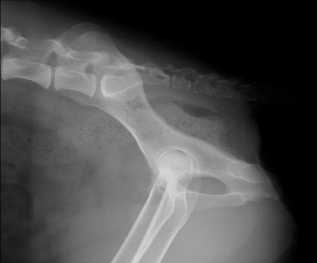
Mediolateral Projection
Click on the circles to see the answer
Crest of Ilium
Sacral Vertebra 3
Promontory of Sacrum
Caudal Vertebra 1
Body of Ilium
Head of the right and left Femurs (superimposed)
Obturator Foramen
Ischiatic Tuberosity
The caudal end of the sacrotuberous ligament attaches to the dorsal surface of this eminence.
The ventral surface of the ischiatic tuberosity gives rise to the largest muscles of the thigh, the hamstring muscles: mm. biceps femoris, semitendinosus, and semimembranosus.

Computed Tomography Anatomy of the Pelvis
Dorsal plane
Click and drag to go through the study. Position your cursor over the structures to see anatomical labels
- Femoral Head
- Femoral Neck
- Acetabulum
- Acetabular Fossa
- Greater Trochanter Lesser Trochanter
- Wing of Ilium
- Body of Ilium
- Pubis
- Ischium
- Wing of Sacrum
3D Reconstruction of the Pelvis
Click and drag to rotate the 3D object
Click on labels to see annotations
Dr Mariano Makara
Dip. ECVDI